Quick answer
High BindingMedium BindingIgG class antibodies ( 150 kda ) at a concentration of 3-5 µg/ml in pH 6-8 buffer, are easily passively adsorbed on both and microplates. When the assay depends on keeping the antigen-binding (Fab)2 arms free, switch to oriented capture: biotinylate the antibody on its Fc region and use a Streptavidin plate or capture the native Fc on a Protein A/G plate or even with a second antibody specific for the native Fc. The method you choose sets the antibody’s orientation, and orientation sets your sensitivity.
Coating an antibody onto a microplate looks like one step in a protocol: dilute, dispense, incubate overnight. The choice underneath that step decides how much of your capture antibody actually works once the antigen arrives. This guide compares the five methods used to immobilize an antibody on a 96-well polystyrene plate, with the binding chemistry, the typical concentration, and the orientation each one produces.
How an antibody binds to an ELISA plate
Antibody immobilization is the attachment of a capture antibody to the well surface so it stays put through every wash and still presents its binding sites to the antigen. There are three ways to make that attachment: physical adsorption, affinity capture, and covalent coupling. For antibodies, the first two cover almost every assay.
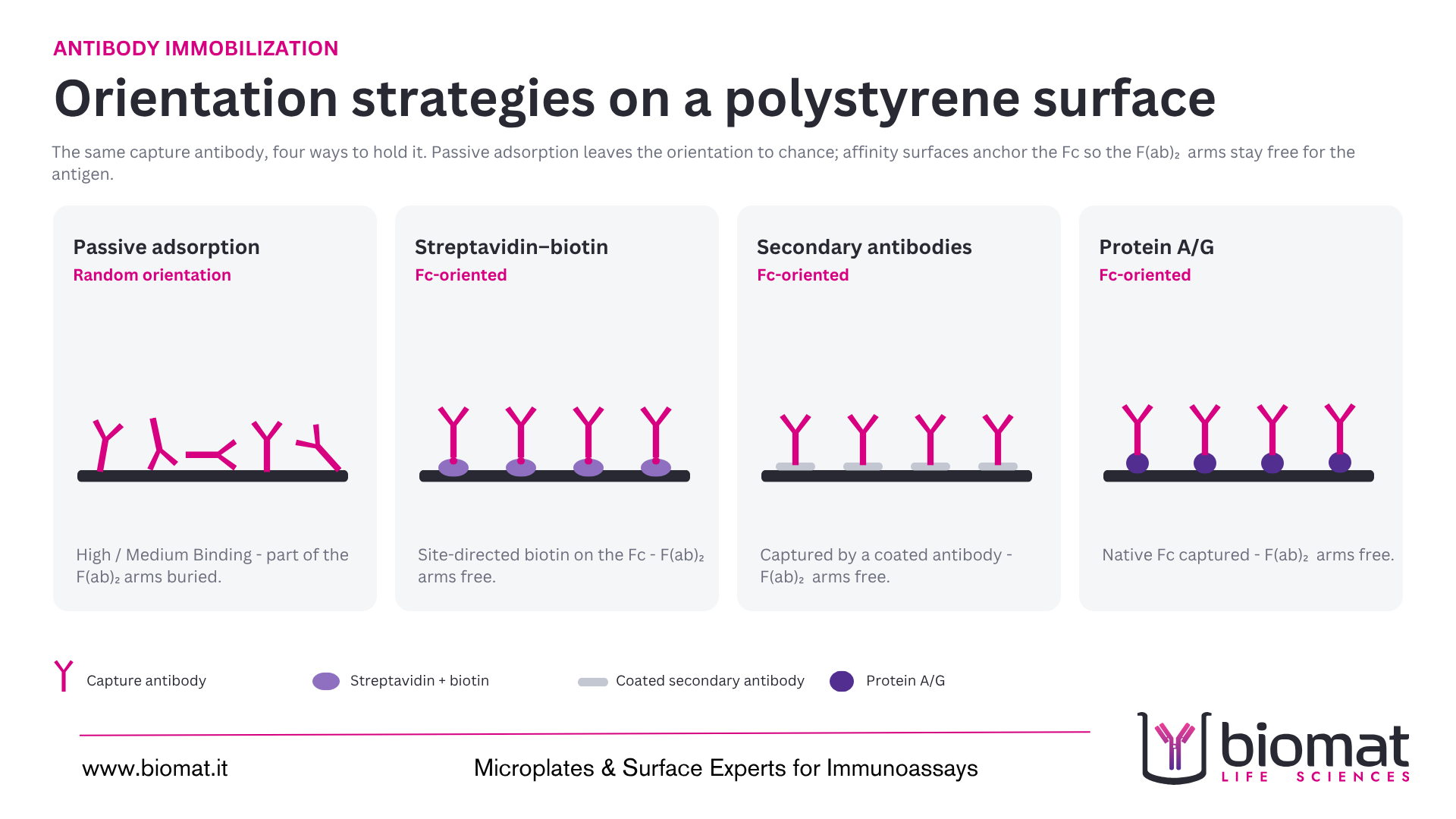
In passive adsorption the antibody sticks to the polystyrene through ionic and hydrophobic interactions. It works, but it lands the antibody in a random orientation: some molecules sit with the F(ab)2 arms exposed, many do not. Affinity capture improves on that, but the degree of improvement depends on where the antibody is held. Protein A/G binds the Fc directly, so the F(ab)2 arms point away from the surface. Streptavidin binds biotin, so the biotinylated antibody, predominantly at its Fc site, is captured, leaving its F(ab)2 group oriented to bind the antigen. A second antibody specific for the Fc of the primary antibody can be coated and thus specifically bind the primary antibody through its Fc, leaving its F(ab)2 free to bind the antigen.
This orientation problem is the single most useful thing to understand before you coat a plate. A surface that binds more antibody is not automatically the surface that gives more signal. What matters is how much functional antibody you immobilize, and that depends on the method.
The five antibody coating methods, compared
The table below matches each method to the antibody type, the recommended pH and post-coating step, the typical surface density, and the orientation you can expect. Values reflect Biomat surface documentation for a 100 µL/well coating volume.
Two footnotes worth keeping in your protocol. A Secondary Antibodies plate carries a defined host species (for example goat anti-mouse IgG or goat anti-rabbit IgG); in a sandwich or direct format the labeled antibody has to come from the matching species. And when you build a sandwich on a Protein A/G plate, the labeled detection antibody should be an F(ab)₂ or F(ab) enzyme conjugate, so the Protein A/G surface does not capture it too.
Match a surface to your antibody. Browse all Biomat 96-well microplates or request a quote with your molecule and detection method and the technical team will narrow it down.
How to choose the method for your antibody
Use this sequence. It mirrors the logic we walk through with assay developers before recommending a surface.
- Is a standard sandwich or competitive assay enough, with a well-behaved antibody? Start with passive adsorption on High Binding. It is the fastest route and needs no antibody modification. For antibodies that give high background on High Binding, move to Medium Binding for a cleaner signal-to-noise ratio.
- Is functional activity dropping because the F(ab)2 arms are buried? This is the classic symptom of random adsorption. Switch to oriented capture. If you can biotinylate the antibody in an Fc-directed way, streptavidin gives you defined, wash-stable orientation. If you would rather not modify the antibody at all, Protein A/G captures the native Fc directly.
- Do you need to capture a specific host species without modifying anything? A Secondary Antibodies surface does that, at the cost of locking you into the matching detection species.
The business side of this choice is concrete, but it is not the same for every oriented method. Streptavidin and secondary-antibody capture often reach the same signal with less antibody (1–1.5 µg/mL versus 3–5 µg/mL for passive adsorption), which lowers reagent cost per plate. Protein A/G is oriented too, but it is coated at a higher concentration (≥4 µg/mL, similar to passive), so there the payoff is orientation and lot-to-lot consistency rather than reagent saving. Either way, a defined orientation tightens reproducibility once you scale a kit into production.
Coating buffer, pH and conditions
Most published ELISA protocol coatings are in carbonate–bicarbonate buffer at pH 9.6, the classic condition for passive adsorption on untreated polystyrene. Biomat surfaces are gamma irradiated and coated in the milder pH 6–8 range, which is gentler on pH-sensitive antibodies and still reaches the binding densities in the table. If you are migrating a protocol optimized at pH 9.6, treat the buffer as a variable to re-check rather than a fixed rule, and titrate on the Biomat surface.
After coating, block with the post-coating step listed in the table to cover the unsaturated surface and cut non-specific binding. General laboratory practice is to incubate overnight at 4 °C or for a shorter period at room temperature, then store coated plates dry and sealed with desiccant. Confirm the exact time, temperature, and shelf life for your antibody experimentally, since these vary with the molecule.
What to do when you require a different surface for your application?
Most capture antibodies are covered by the five methods above. If the above-mentioned attack methods do not work you could interface with Biomat.
Biomat controls treatment and coating in-house and runs pilot batches for custom surfaces, typically 10-20 plates for validation before production volumes. If the table above does not point to an obvious answer for your antibody, that is usually the signal for a tailored surface.
For the full decision logic across all biomolecule types, see the microplate surface selection guide. To match a surface to a specific antibody and assay, request a quote with your molecule and detection method, and the technical team will narrow it down.
Not sure which microplates surface best fits you needs?
Talk to our R&D team
References
- Engvall E, Perlmann P. Enzyme-linked immunosorbent assay (ELISA). Quantitative assay of immunoglobulin G. Immunochemistry, 1971. (Foundational ELISA method – link at publish, target=”_blank” rel=”noopener”.)
- Hermanson GT. Bioconjugate Techniques, 3rd ed. Academic Press. (Standard reference for biotinylation, EDC/amine and maleimide coupling chemistry – link at publish.)
Biomat is a specialist in surface modification and custom plate engineering, delivering high-performance microplates and tailored plastic consumables for demanding diagnostic and research applications. Produced under an ISO 9001:2015 quality management system. Made in Italy, since 1994.